ERcast: Clinical Perspectives Podcast Preview
The summary below is from an episode of ERcast: Clinical Perspectives
Multiple myeloma often first declares itself in the ED through organ injury rather than a known cancer diagnosis. CRAB findings—hypercalcemia, renal dysfunction, anemia, and bone lesions—should sharpen suspicion, especially when anemia, acute kidney injury, and hypercalcemia cluster together.
Recognizing Multiple Myeloma in the ED
- CRAB complication pattern: Hypercalcemia, renal dysfunction, anemia, and bone lesions are the classic bedside clues, and the combination of anemia with acute kidney injury or hypercalcemia should prompt a myeloma workup.
- Typical initial presentations: Unexplained anemia is the most common presenting feature, with bony pain, acute kidney injury, fatigue, weight loss, and hypercalcemia rounding out the usual emergency-department picture.
- High-yield ED testing: CBC, BMP, and ionized calcium are the key emergency tests, with ionized calcium being unusually helpful here because total calcium can mislead in clinically important myeloma hypercalcemia.
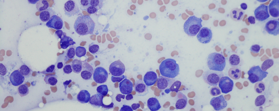
- Definitive diagnosis pathway: Urine protein electrophoresis and bone marrow biopsy establish the diagnosis, but the emergency task is recognizing the complication pattern early enough to trigger the right downstream evaluation. We get into that diagnostic handoff in the episode.
- Imaging for bone pain: Plain radiographs are the starting point for focal bony pain, while CT can better define lytic lesions and structural risk when a pathologic process is already on the table.
Major Myeloma Complications and Management
- Renal injury burden: Acute kidney injury is common, with creatinine often already above 2 mg/dL at diagnosis, and supportive care matters because kidney dysfunction is reversible in a meaningful share of patients.
- Hypercalcemia framing: Myeloma hypercalcemia is driven by bone demineralization, and severe cases are treated with IV fluids, calcitonin, and a bisphosphonate, with dialysis sometimes needed for the sickest patients.
- Infection risk window: Infection risk is markedly elevated—about 7- to 11-fold higher than the general population—and is highest early in treatment, so sepsis coverage should be broad and early, with antivirals added when viral disease is plausible.
- Skeletal instability clues: Osteolytic disease is present in roughly 70% of patients, and pathologic or insufficiency fractures are common enough that any fracture in suspected myeloma deserves a more careful imaging strategy. We walk through when to escalate imaging in the chapter.
- Spinal cord red flags: Nerve root compression is the most important neurologic complication, and cauda equina syndrome makes MRI and urgent specialty involvement time-critical.
- Therapy-related thrombosis: Myeloma carries a strikingly elevated VTE risk, especially in the first 6 months of therapy, and confirmed DVT or PE is generally treated with LMWH or a DOAC.
Subscribe to ERcast: Clinical Perspectives to listen to the episode.
References:
- Long B, McCurdy A, Koyfman A, Rosenberg H. An emergency medicine review: Multiple myeloma and its complications. Am J Emerg Med. 2025 Feb;88:172-179. PMID: 39643958.
Faculty
- Drew Kalnow, DO
Dr. Drew Kalnow is an emergency medicine physician and educator based in Columbus, Ohio. He completed his emergency medicine training at OhioHealth Doctors Hospital Emergency Medicine Residency. Dr. Kalnow is passionate about advancing emergency medicine through high-quality education, with a particular focus on simulation, learning theory, and innovative teaching.
- Brit Long, MD
Dr. Brit Long is a Professor of Emergency Medicine at the University of Virginia and an emergency medicine physician with experience in both a community ED and at a military academic center ED. He is the Clinical Editor-in-Chief of emDOCs.His professional interests include medical education, evidence-based medicine, and the FOAMed movement. Outside of work, he enjoys spending time with his wife and two daughters