ERcast: Clinical Perspectives Podcast Preview
The summary below is from an episode of ERcast: Clinical Perspectives
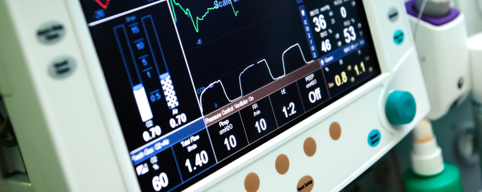
Capnography is more than an endotracheal tube check: end-tidal CO2 reflects CO2 production, perfusion, and ventilation across the whole body. In the ED, waveform capnography can flag hypoventilation before pulse oximetry and add useful bedside signal in cardiac arrest, DKA, sepsis, trauma, PE, and obstructive lung disease.
Capnography Fundamentals in the ED
- Whole-body CO2 signal: End-tidal CO2 is a systems-level marker of metabolism, circulation, and ventilation, so the number only makes sense when you interpret it alongside the waveform and the clinical context.
- Normal EtCO2 range: A typical end-tidal CO2 range is 35-45 mm Hg, but bedside interpretation hinges just as much on capnogram shape and the arterial-to-end tidal CO2 gradient.
- Waveform phase recognition: Phase III is the alveolar plateau, and its height and slope often carry the most clinical information when you are looking for bronchospasm, hypoventilation, or worsening dead space.
- Faster than pulse oximetry: Capnography detects hypoventilation earlier than pulse oximetry, making respiratory depression visible before oxygen saturation starts to fall. We get into the practical bedside implications in the episode.
- Important technical confounders: Ventilator problems, tubing leaks, obstruction, disconnection, and monitor malfunction can all distort EtCO2, so a surprising value should trigger a quick equipment check before overcalling pathology.
High-Yield Clinical Uses and Limits
- Airway confirmation standard: Quantitative waveform capnography is more reliable than fogging, chest rise, or breath sounds for confirming tracheal intubation and for catching tube displacement during transport.
- Cardiac arrest perfusion marker: During arrest, EtCO2 tracks cardiopulmonary blood flow and compression quality; a sudden rise of at least 10 mm Hg is highly specific for ROSC.
- DKA bedside discriminator: In hyperglycemic patients, an EtCO2 above 35 mm Hg is highly sensitive for ruling out DKA, while values below 24 mm Hg strongly support the diagnosis.
- Trauma and shock signal: Low EtCO2 correlates with injury severity, transfusion need, and mortality in trauma, and a 2 mm Hg rise with straight-leg raise can suggest fluid responsiveness.
- Obstructive airway waveform: Bronchospasm produces the classic shark-fin capnogram as Phase III steepens, and serial waveform improvement can mirror response to bronchodilators and steroids.
- Adjunct not answer key: Capnography works best when one dominant physiology is driving the picture; mixed shock states, low perfusion, and rare false positives can mislead if the tracing is read in isolation. We walk through the practical limitations in the chapter.
Subscribe to ERcast: Clinical Perspectives to listen to the episode.
References:
- Long B, Koyfman A, Vivirito MA. Capnography in the Emergency Department: A Review of Uses, Waveforms, and Limitations. J Emerg Med. 2017;53(6):829-842. PMID: 28993038
- Manifold CA, Davids N, Villers LC, Wampler DA. Capnography for the nonintubated patient in the emergency setting. J Emerg Med. 2013;45(4):626-632. PMID: 23871325
- Nassar BS, Schmidt GA. Capnography During Critical Illness. Chest. 2016;149(2):576-585. PMID: 26447854
- Godwin SA, Burton JH, Gerardo CJ, et al. Correction: Correction to 'Clinical Policy: Procedural Sedation and Analgesia in the Emergency Department' [Annals of Emergency Medicine 63 (2014) 247-258.e18]. Ann Emerg Med. 2017;70(5):758. PMID: 28395927
- Panchal AR, Berg KM, Hirsch KG, et al. 2019 American Heart Association Focused Update on Advanced Cardiovascular Life Support: Use of Advanced Airways, Vasopressors, and Extracorporeal Cardiopulmonary Resuscitation During Cardiac Arrest: An Update to the American Heart Association Guidelines for Cardiopulmonary Resuscitation and Emergency Cardiovascular Care. Circulation. 2019;140(24):e881-e894. PMID: 31722552
- Soleimanpour H, Taghizadieh A, Niafar M, Rahmani F, Golzari SE, Esfanjani RM. Predictive value of capnography for suspected diabetic ketoacidosis in the emergency department. West J Emerg Med. 2013;14(6):590-594. PMID: 24381677
- Bou Chebl R, Madden B, Belsky J, Harmouche E, Yessayan L. Diagnostic value of end tidal capnography in patients with hyperglycemia in the emergency department. BMC Emerg Med. 2016;16:7. Published 2016 Jan 29. PMID: 26821648
- Hemnes AR, Newman AL, Rosenbaum B, et al. Bedside end-tidal CO2 tension as a screening tool to exclude pulmonary embolism. Eur Respir J. 2010;35(4):735-741. PMID: 19717480
- Riaz I, Jacob B. Pulmonary embolism in Bradford, UK: role of end-tidal CO2 as a screening tool. Clin Med (Lond). 2014;14(2):128-133. PMID: 24715122
Faculty
- Brit Long, MD
Dr. Brit Long is a Professor of Emergency Medicine at the University of Virginia and an emergency medicine physician with experience in both a community ED and at a military academic center ED. He is the Clinical Editor-in-Chief of emDOCs.His professional interests include medical education, evidence-based medicine, and the FOAMed movement. Outside of work, he enjoys spending time with his wife and two daughters
- Jenny Beck-Esmay, MD