ERcast: Clinical Perspectives Podcast Preview


The summary below is from an episode of ERcast: Clinical Perspectives
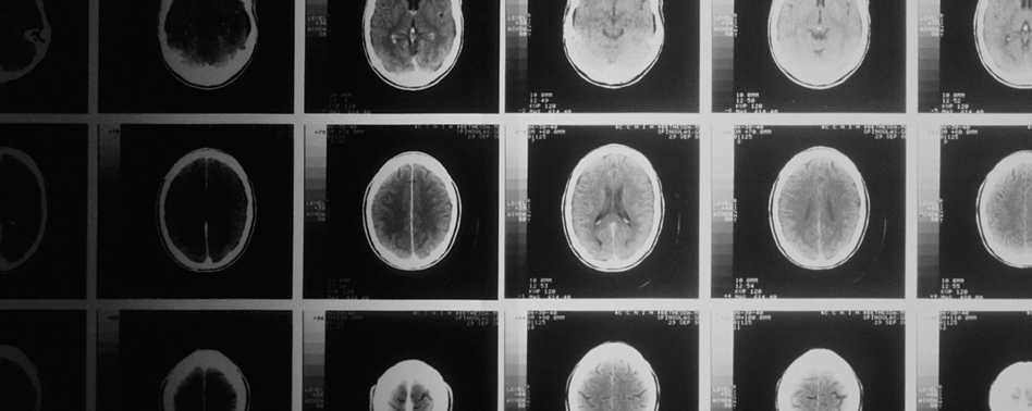
Time-sensitive intracranial hemorrhage, mass effect, hydrocephalus, and herniation can often be recognized on noncontrast head CT before a formal radiology read returns. A structured emergency medicine approach starts with blood, then cisterns, brain, ventricles, vessels, and bone.
Systematic Head CT Life Threats
- Pretest probability first: High-risk patients with a strong pretest probability of a time-dependent intracranial lesion should not wait passively for teleradiology, because clinically important delays and meaningful discrepancy rates are real.
- Blood then location: Acute intracranial blood is typically bright white on CT once clotted, and the first job is to decide whether blood is present, where it sits, and whether it is causing shift or compression.
- Cistern compression sign: Compressed basal cisterns are an early imaging clue to rising intracranial pressure, so checking whether the key cisterns are open may matter before more dramatic herniation signs appear. We walk through the named cisterns in the episode.
- Brain symmetry check: Normal brain parenchyma should look symmetric with preserved gray-white differentiation and an intact cortical gyral pattern; asymmetry, edema, mass effect, or midline shift signals danger.
- Hydrocephalus early clue: The ventricles should be dark and slitlike rather than ballooned, and enlargement of the temporal horns is an early clue to hydrocephalus before more obvious ventricular dilation appears.
- Hyperdense vessel warning: On a noncontrast head CT, a vessel that looks conspicuously dense usually means clot rather than flowing blood, making the hyperdense vessel sign a useful emergency red flag.
Bone Windows and Practical Pearls
- Bone versus blood: Bone and calcified choroid can also appear white, but blood is far less dense than bone on Hounsfield units, a practical distinction when a bright focus is not in an expected location.
- Basilar fracture clue: Mastoid air cells should be filled with air; opacified mastoids after head trauma mean blood until proven otherwise and should raise concern for basilar skull fracture.
- Use bone windows: Skull fracture detection improves when you window the scan for bone rather than trying to judge fractures on soft-tissue settings, a simple adjustment that changes what you can reliably see.
- Systematic normal scan: A reassuring noncontrast head CT has no blood, open cisterns, symmetric brain with preserved gray-white differentiation, no ventricular dilation, no hyperdense vessels, and no fracture. That rapid sweep is worth hearing in the chapter.
Subscribe to ERcast: Clinical Perspectives to listen to the episode.
References:
- Kennedy S, et al. The effect of teleradiology on time to interpretation for CT pulmonary angiographic studies. J Am Coll Radiol. 2009 Mar;6(3):180-189.e1. PMID: 19248994.
- Platts-Mills TF, Hendey GW, Ferguson B. Teleradiology interpretations of emergency department computed tomography scans. J Emerg Med. 2010 Feb;38(2):188-95. PMID: 18814996.
- Storjohann S, et al. The Accuracy of On-Call CT Reporting in Teleradiology Networks in Comparison to In-House Reporting. Healthcare (Basel). 2021 Apr 1;9(4):405. PMID: 33916229.
- Walker EA, et al. Quality metrics currently used in academic radiology departments: results of the QUALMET survey. Br J Radiol. 2017 Mar;90(1071):20160827. Link.
- Jamal K, et al. 'Out of hours' adult CT head interpretation by senior emergency department staff following an intensive teaching session: a prospective blinded pilot study of 405 patients. Emerg Med J. 2014 Jun;31(6):467-70. PMID: 23576233.
Faculty
- Matthew DeLaney, MD, FACEP, FAAEM
Dr. Matthew DeLaney is an emergency medicine physician and educator based in Birmingham, Alabama. A native of Mobile, he earned his medical degree from the University of South Alabama and completed his emergency medicine residency at Maine Medical Center.Dr. DeLaney has experience in both community and academic emergency medicine and is known for his commitment to teaching and medical education. He lives in Birmingham with his wife, Erin, who is also a physician, and their two daughters.
- Andrew Perron, MD