ERcast: Clinical Perspectives Podcast Preview



Hippo ERcast April 2022
- Apr 2022
- 7 Chapters
- 2 hr 33 min
Faculty
- Matthew DeLaney, MD, FACEP, FAAEM
Dr. Matthew DeLaney is an emergency medicine physician and educator based in Birmingham, Alabama. A native of Mobile, he earned his medical degree from the University of South Alabama and completed his emergency medicine residency at Maine Medical Center.Dr. DeLaney has experience in both community and academic emergency medicine and is known for his commitment to teaching and medical education. He lives in Birmingham with his wife, Erin, who is also a physician, and their two daughters.
- Rob Orman, MD
Dr. Rob Orman is an emergency physician, educator, and executive coach specializing in physician performance and professional fulfillment. After more than 20 years in community emergency medicine, he now works with clinicians across specialties to address burnout, inefficiency, and career challenges. He earned his medical degree from Emory University School of Medicine and completed his residency at Denver Health Medical Center, where he served as chief resident. Dr. Orman is the founder of the Stimulus podcast and Orman Physician Coaching. He previously served as chief editor of ERcast and hosted Essentials of Emergency Medicine for nearly a decade.
- Drew Kalnow, DO
Dr. Drew Kalnow is an emergency medicine physician and educator based in Columbus, Ohio. He completed his emergency medicine training at OhioHealth Doctors Hospital Emergency Medicine Residency. Dr. Kalnow is passionate about advancing emergency medicine through high-quality education, with a particular focus on simulation, learning theory, and innovative teaching.
- Charles Khoury MD, FACEP, FAAEM
- Kristin Crichton, MD
- Andrew Perron, MD
- Arun Sayal, MD



Chapters
Drew Kalnow on SVC syndrome
Superior vena cava syndrome can present like a COPD exacerbation, but facial and neck edema with intermittent stridor should shift concern toward obstructive airway disease. Malignancy and catheter-related thrombosis now drive most cases, and early airway planning matters more than reassuring initial appearance. SVC Syndrome Recognition and Airway Misleading COPD mimic: Wheezing with oxygen saturations in the 80s can distract from the real problem; upper torso, neck, and facial edema out of proportion to the rest of the body is the bedside clue. Modern etiologic pattern: Most SVC syndrome now comes from malignancy or central-line-associated thrombosis rather than infection, and that split determines whether you are thinking tumor therapy, thrombolysis, or stenting. Obstructive airway warning: Intermittent stridor in SVC syndrome is a red flag for airway edema or tracheal compression, even when the patient is still talking and initially mentating well. Early airway strategy: Awake intubation is the safer default when edema threatens the airway, and paralytics can turn a tenuous situation into a cannot-intubate problem. We get into the backup planning in the episode. Rescue temporizing measures: Racemic epinephrine and steroids may shrink tissue edema enough to buy time, but they do not fix the underlying obstruction and should not delay definitive airway or specialty help. Etiology-directed treatment: Malignant SVC syndrome often pushes you toward steroids and careful decongestion, while severe thrombotic disease may need thrombolysis with vascular or IR involvement for possible stenting.
EEM Rewind: A metatarsal, a Jones, and a Lisfranc walk into a bar
Fifth metatarsal fractures are common, but not all lateral foot injuries are benign. Jones fractures carry a meaningful nonunion risk, and Lisfranc injuries are the classic missed medial midfoot injury after a twisting plantar-flexion mechanism. Metatarsal fractures in the ED Base fifth metatarsal fractures: Tuberosity avulsion fractures are the most common fifth metatarsal injury and usually do well with a stiff shoe or walking boot plus weight bearing as tolerated. Jones fracture risk: A true proximal fifth metatarsal fracture occurs distal to the tuberosity and has a 10-15% nonunion risk because the blood supply is poor in that zone. Shaft and neck patterns: More distal fifth metatarsal shaft or neck fractures are often oblique and generally heal nonoperatively, though recovery commonly takes a couple of months. Central metatarsal stability: Isolated low-energy fractures of the second through fourth metatarsals are usually stable enough for a walking boot, while multiple or high-energy injuries deserve more protection. Stress fracture caveat: Second, third, and fourth metatarsal stress fractures can have a normal initial x-ray, so persistent focal pain after overuse still warrants immobilization and follow-up. We get into the practical splinting choices in the episode. Lisfranc injuries and missed midfoot trauma Classic injury mechanism: Lisfranc injury follows a twisting force on a plantar-flexed, externally rotated foot, the setup seen after stepping in a hole or landing awkwardly. Medial midfoot red flag: Inability to bear weight with medial midfoot pain and tenderness after a twisting injury should be treated as Lisfranc until proven otherwise. Subtle radiographic clues: Plain films may look normal, but malalignment at the first or second tarsometatarsal joints and a tiny avulsion fleck sign are classic clues. Limits of advanced imaging: CT helps define bony injury for operative planning, but purely ligamentous Lisfranc injuries can still be missed, so the exam matters most. That nuance is worth hearing in the chapter. Initial ED disposition: Suspected or minimally displaced Lisfranc injuries need a posterior slab, non-weight bearing, and close orthopedic follow-up, while dislocation requires urgent reduction planning.
Non-Accidental Trauma
Non-accidental trauma is a pattern-recognition diagnosis anchored in developmental age, injury distribution, and occult injury screening. In infants and young children, bruising in soft-tissue locations, unexplained vomiting or altered mental status, and fractures out of proportion to the history should trigger a child-abuse workup and mandatory reporting review. Recognizing and Working Up NAT Developmental age mismatch: Mechanism only makes sense in the context of what the child can actually do; a history like a young infant “rolling off” furniture should immediately raise concern for inflicted injury. Bruising pattern red flags: Bruises over bony prominences can be accidental, but bruising on softer areas or across the body is far less reassuring and should push abuse higher on the differential. Complete exposed exam: The initial evaluation starts with fully undressing the child, a full skin exam including around the ears, and careful inspection of the oropharynx for a torn frenulum or other hidden trauma. Skeletal survey yield: Skeletal survey is a core test in suspected abuse, especially in children under 2 years or with developmental delay, because occult fractures are often missed on routine exam. Head injury screening: Head CT matters early because abusive head trauma can be clinically subtle; get neuroimaging for altered mental status, unexplained vomiting, facial trauma, and any concerned infant under 7 months. We walk through the imaging triggers in the episode. Abdominal injury labs: AST, ALT, and lipase screen for occult abdominal trauma in younger children, with abnormal results prompting CT imaging, while CBC and coagulation studies help assess bleeding and important mimics. Reporting and Family Interaction Partner with the caretaker: Start from the shared goal of the child’s safety and health; the adult who brought the child in may not be the perpetrator, so avoid turning the bedside interview into an investigation. Trust your concern signal: If the story, exam, or injury pattern feels off, acknowledge that instinct and act on it; early concern is often what prevents missed abuse in preverbal children. Mandated reporting threshold: The safest approach is to report any reasonable suspicion of abuse or neglect, and transfer to another hospital does not remove that obligation. We get into the reporting nuances in the episode. Law enforcement involvement: Involve law enforcement urgently when a parent or guardian refuses transport or evaluation despite safety concerns, because immediate scene control may be necessary to protect the child. Reporting practicalities: Know your local statute, report promptly, and answer unknowns honestly; “I do not know” is appropriate when details are unclear and should not delay contacting child protective services. Discipline versus abuse: Physical discipline that leaves a mark is abuse and reportable; distinguishing accepted discipline from abuse becomes straightforward once there is visible injury.
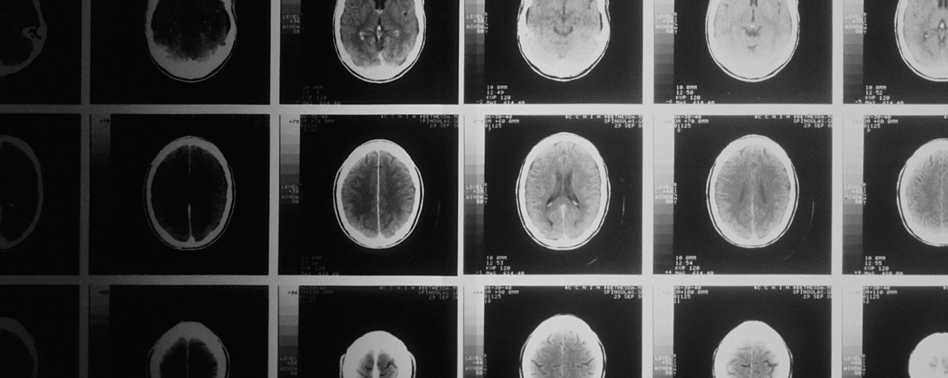
Head CT’s: Blood Can Be Very Bad
Time-sensitive intracranial hemorrhage, mass effect, hydrocephalus, and herniation can often be recognized on noncontrast head CT before a formal radiology read returns. A structured emergency medicine approach starts with blood, then cisterns, brain, ventricles, vessels, and bone. Systematic Head CT Life Threats Pretest probability first: High-risk patients with a strong pretest probability of a time-dependent intracranial lesion should not wait passively for teleradiology, because clinically important delays and meaningful discrepancy rates are real. Blood then location: Acute intracranial blood is typically bright white on CT once clotted, and the first job is to decide whether blood is present, where it sits, and whether it is causing shift or compression. Cistern compression sign: Compressed basal cisterns are an early imaging clue to rising intracranial pressure, so checking whether the key cisterns are open may matter before more dramatic herniation signs appear. We walk through the named cisterns in the episode. Brain symmetry check: Normal brain parenchyma should look symmetric with preserved gray-white differentiation and an intact cortical gyral pattern; asymmetry, edema, mass effect, or midline shift signals danger. Hydrocephalus early clue: The ventricles should be dark and slitlike rather than ballooned, and enlargement of the temporal horns is an early clue to hydrocephalus before more obvious ventricular dilation appears. Hyperdense vessel warning: On a noncontrast head CT, a vessel that looks conspicuously dense usually means clot rather than flowing blood, making the hyperdense vessel sign a useful emergency red flag. Bone Windows and Practical Pearls Bone versus blood: Bone and calcified choroid can also appear white, but blood is far less dense than bone on Hounsfield units, a practical distinction when a bright focus is not in an expected location. Basilar fracture clue: Mastoid air cells should be filled with air; opacified mastoids after head trauma mean blood until proven otherwise and should raise concern for basilar skull fracture. Use bone windows: Skull fracture detection improves when you window the scan for bone rather than trying to judge fractures on soft-tissue settings, a simple adjustment that changes what you can reliably see. Systematic normal scan: A reassuring noncontrast head CT has no blood, open cisterns, symmetric brain with preserved gray-white differentiation, no ventricular dilation, no hyperdense vessels, and no fracture. That rapid sweep is worth hearing in the chapter.

Lit Matters #1: High vs. Low Dose IV Insulin for Asymptomatic Hyperglycemia
Asymptomatic hyperglycemia in the ED rarely needs aggressive IV insulin just to improve a discharge glucose. In non-emergent hyperglycemia, giving more than 5 units IV lowered glucose more than 5 units or less, but did not shorten ED length of stay and still carried a meaningful hypokalemia signal. IV Insulin for ED Hyperglycemia Non-emergent hyperglycemia framing: Asymptomatic hyperglycemia is not DKA, and the key management question is clinical benefit rather than how fast the number falls. Dose comparison cutoff: Using more than 5 units of IV regular insulin produced a larger glucose drop than 5 units or less, but that extra reduction did not translate into a shorter ED stay. Length of stay reality: Median ED length of stay was 240 minutes in both groups, a practical reminder that pushing the glucose down faster does not necessarily move disposition. Electrolyte downside: Hypokalemia showed up in about 8% of patients despite this being a non-emergent population, which makes IV regular insulin less benign than it often feels at the bedside. Fluids and renal function: More than 2 liters of IV fluids tracked with bigger glucose reduction, while better renal function tracked with a smaller drop. We get into why that matters for bedside decision-making in the episode. Practice goal check: The real question is what you are trying to accomplish before discharge, because a lower glucose number alone did not produce a measurable operational benefit.

Lit Matters #2: The Impact of Availability Bias on ED Workups
Availability bias measurably changes emergency department workups for shortness of breath. After an ED physician diagnoses pulmonary embolism, PE testing in subsequent dyspnea patients rises transiently, even in a setting with decision rules and relatively lower medicolegal pressure. Availability Bias in PE Workups Transient post-PE testing spike: A recent pulmonary embolism diagnosis nudges the next dyspnea workups toward more D-dimer and CTPA use, with about a 15% relative increase in PE testing over the next 10 days. Signal despite decision rules: This effect showed up in PE, a diagnosis already supported by tools like Wells and PERC, suggesting cognitive bias persists even when structured risk stratification exists. Short-lived recency effect: The testing increase was not permanent; it drifted back toward baseline over the following weeks, a practical reminder that recent memorable cases can temporarily reshape pretest probability. Built-in negative controls: The study compared PE testing changes with unrelated orders like TSH and hand radiographs, strengthening the case that the shift reflected availability bias rather than a general tendency to overtest. Why this likely matters more: A VA-based cohort may underestimate the real-world effect, since broader ED practice includes more medicolegal pressure and additional PE risk contexts. We get into the bedside implications in the episode.

Lit Matters #3: Rethinking the Time of HA Onset to CT Cutoff for SAH
Modern multislice noncontrast head CT appears highly sensitive for aneurysmal subarachnoid hemorrhage beyond the classic 6-hour window, but the bedside meaning is still unsettled. The real question is not just missed blood on CT, but whether a missed SAH would change management or require neurosurgical intervention. Modern CT for SAH Timing Twenty-four hour sensitivity signal: In this 10-year single-center analysis, modern multislice CT identified all aneurysmal SAH cases within 24 hours of headache onset, challenging the traditional 6-hour rule-out boundary. Intervention-focused miss risk: Among patients scanned within 24 hours, the clinically important signal was that no CT-missed SAH went on to require a neurosurgical procedure, a distinction worth hearing in the episode. Retrospective design limits: The study worked backward from known SAH cases, which raises spectrum and ascertainment concerns and makes the reassuring sensitivity estimate less ready for broad bedside adoption. Imputed onset uncertainty: Headache-onset time was missing in 224 of 347 cases, so the authors used best-case and worst-case imputation rather than observed timing, a major caveat for any time-based cutoff. Confidence interval reality: The reported 24-hour sensitivity for aneurysmal SAH carried a 95% confidence interval that did not prove perfection, which matters when the disease in question can be catastrophic or fatal. Technology versus implementation: Third-generation multislice scanners performed similarly across 64- and 128-slice platforms, suggesting scanner era alone is not the whole story behind safe SAH exclusion.