ERcast: Clinical Perspectives Podcast Preview
The summary below is from an episode of ERcast: Clinical Perspectives
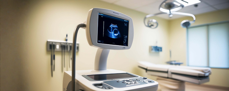
Point-of-care ultrasound can rapidly narrow the differential in undifferentiated shock and identify immediately reversible causes of arrest. In resuscitation, its highest value is as a bedside physiologic data point for cardiac function, tamponade, right-heart strain, pneumothorax, and volume tolerance.
Ultrasound for Shock and Arrest
- Undifferentiated shock framing: POCUS is most useful when shock is clinically unclear, helping separate cardiogenic, obstructive, and hemorrhagic patterns quickly enough to change the next test, consultant, and first moves.
- Gross cardiac function check: A rapid look at overall myocardial squeeze is an essential bedside skill in crashing patients, and often gives a faster hemodynamic read than waiting for formal imaging.
- Femoral flow assessment: Femoral artery flow on ultrasound can be more informative than pulse palpation during a code, especially when pulse checks are equivocal. We get into the bedside technique in the episode.
- Myocardial motion in arrest: True myocardial motion matters more than blood swirling in the chambers and is a stronger survival prognostic sign than many clinicians realize.
- Standstill prognostic caution: Cardiac standstill carries a grim prognosis, but it is not absolute; rare patients still survive, so ultrasound findings have to be interpreted in the full clinical context.
- Shockable rhythm limitation: In clearly shockable arrests, ultrasound often adds less because the immediate priority is defibrillation, stabilization, and rapid cath-lab thinking rather than image-driven diagnosis.
Focused Resuscitation Ultrasound Exam
- Core multiorgan survey: A high-yield resuscitation scan pairs the heart with IVC, lungs, and FAST views to look for tamponade, RV enlargement, free fluid, pleural pathology, and fluid tolerance clues.
- Right ventricle clues: An enlarged right ventricle in the right context raises concern for obstructive shock from pulmonary embolism, especially when paired with the rest of the resuscitation exam.
- IVC interpretation limits: IVC size and collapsibility can support fluid decisions, but they are not standalone truth and should never override the broader hemodynamic picture.
- Lung ultrasound yield: Lung views can quickly identify pneumothorax, pleural effusion, and B-lines, giving actionable information during shock without moving an unstable patient.
- Optional DVT extension: A focused DVT study can complement a PE workup when the cardiac views suggest right-heart strain. We walk through when that add-on scan is worth the time in the chapter.
- Outcome reality check: Ultrasound improves diagnostic certainty and resuscitation direction more reliably than mortality, so its value is in sharpening bedside decisions rather than acting as a magic test.
Subscribe to ERcast: Clinical Perspectives to listen to the episode.
References:
- Kim DJ, et al. POCUS literature primer: key papers on POCUS in cardiac arrest and shock. CJEM. 2024 Jan;26(1):15-22. Epub 2023 Nov 23. PMID: 37996693.
- Jones AE, et al. Randomized, controlled trial of immediate versus delayed goal-directed ultrasound to identify the cause of nontraumatic hypotension in emergency department patients. Crit Care Med. 2004 Aug;32(8):1703-8. PMID: 15286547.
- Shokoohi H, et al. Bedside Ultrasound Reduces Diagnostic Uncertainty and Guides Resuscitation in Patients With Undifferentiated Hypotension. Crit Care Med. 2015 Dec;43(12):2562-9. PMID: 26575653.
- Atkinson PR, et al. Does Point-of-Care Ultrasonography Improve Clinical Outcomes in Emergency Department Patients With Undifferentiated Hypotension? An International Randomized Controlled Trial From the SHoC-ED Investigators. Ann Emerg Med. 2018 Oct;72(4):478-489. PMID: 29866583.
- Díaz-Gómez JL, Mayo PH, Koenig SJ. Point-of-Care Ultrasonography. N Engl J Med. 2021 Oct 21;385(17):1593-1602. PMID: 34670045.
Faculty
- Matthew DeLaney, MD, FACEP, FAAEM
Dr. Matthew DeLaney is an emergency medicine physician and educator based in Birmingham, Alabama. A native of Mobile, he earned his medical degree from the University of South Alabama and completed his emergency medicine residency at Maine Medical Center.Dr. DeLaney has experience in both community and academic emergency medicine and is known for his commitment to teaching and medical education. He lives in Birmingham with his wife, Erin, who is also a physician, and their two daughters.
- Andrew Fried, MD
